


When you go to the dentist, after a visual examination of the mouth, the doctor often suggests taking X-rays to complete the diagnosis.
I often hear people say, “I had a panoramic x-ray, so I already have all the necessary documentation.” Not to mention the fact that in the now widespread low-cost clinics, panoramic x-rays are always performed immediately on all patients, without any distinction.
Well, panoramic x-rays often turn out to be USELESS for many types of dental problems.
But how my dentist always only had me do panoramics!!!
Well… what can I say, I’d change him. Without a doubt.
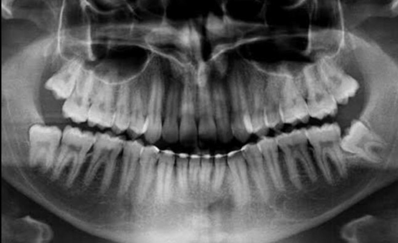 A panoramic X-ray is a highly distorted image and is absolutely not suitable for diagnosing tooth decay or root problems such as granulomas.
It is used for a comprehensive assessment of orthodontic treatment plans (if braces are needed) or extraction surgery.
Especially for extraction surgery of impacted wisdom teeth, it is often combined with a cone beam CT scan, which allows for a three-dimensional assessment of the position of the teeth in relation to important anatomical structures, such as the mandibular nerve.
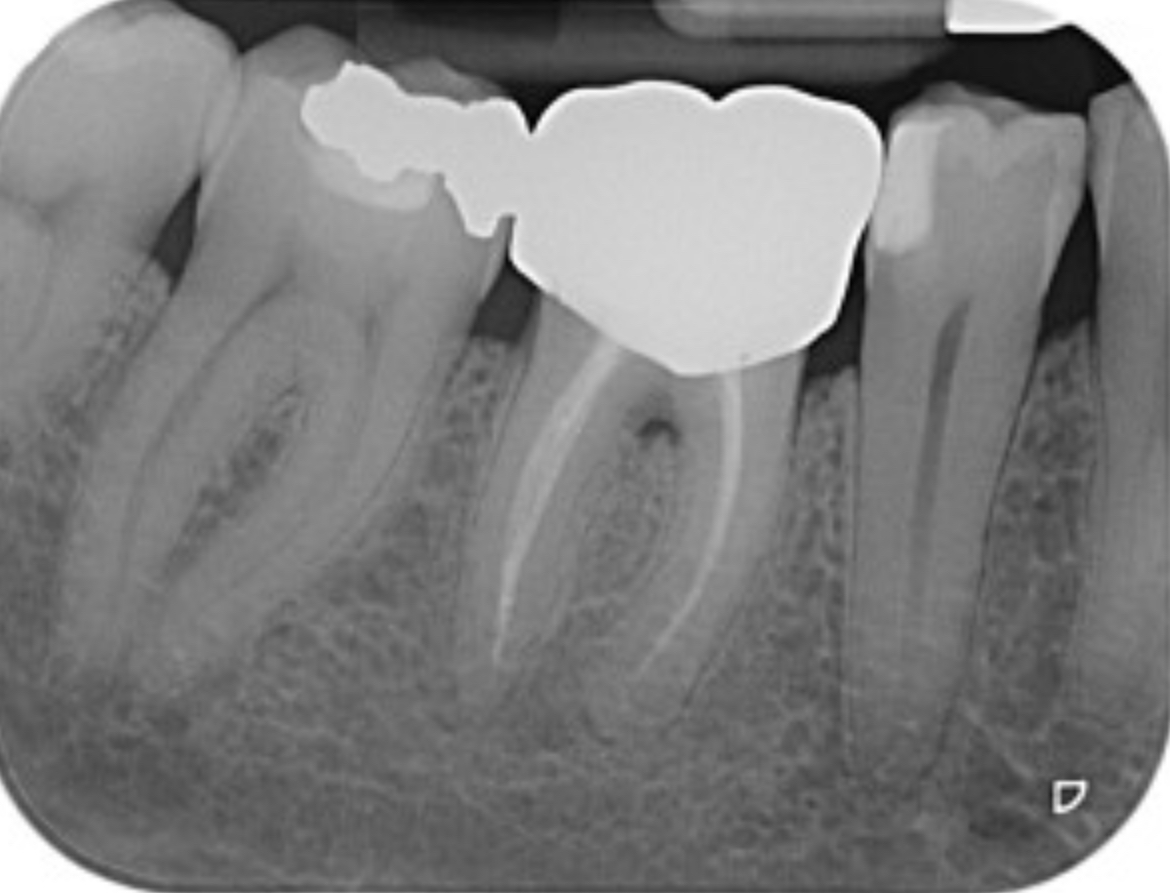 Intraoral x-rays (X-rays) are a valuable aid in diagnosing caries, root lesions, bone lesions, and lesions due to periodontal disease (pyorrhea).
These X-rays reveal a wealth of detail, are reproducible in their position, and therefore comparable; they are essential for assessing the progressive healing of root lesions or for monitoring the progression of periodontal disease over time by monitoring the bone level.
Over the years, significant progress has been made to obtain better-quality and higher-definition images, minimizing the required radiation dose.
How much radiation do I get? Is there a health risk?
The radiation that allows us to obtain x-rays is ionizing, therefore theoretically not zero impact on our organism. But only the area affected by the positioning of the X-ray is directly hit by the rays, which are very sharp and by law in dental X-rays there are constant checks by qualified experts, to protect patients and operators, therefore theoretically not zero impact on our organism. Furthermore, today all the X-rays we use are digital so they have a very low radiation dose.
To get an idea, an intraoral X-ray exposes you like an hour on an airliner, where you are exposed to the sun’s radiation which at high altitudes is less filtered by the atmosphere.
However, it is always necessary to consider the cost-benefit ratio and therefore the important diagnostic information that can be collected.
 In some cases, generally surgical and for the placement of dental implants, a CT scan is necessary. The latest innovation in this field is cone beam computed tomography, which requires a significantly reduced radiation dose and is ideal for use in dentistry.
But how often do I have to do the x-rays?
The evaluation is up to the dentist, the literature suggests a set of x-rays every six months.
My patients generally have hygiene boosters and check-ups every 4 or 6 months and have x-rays once a year.
In this way it is easy to carry out good prevention which is the only truly effective tool in our hands, to avoid having to resort to continuous treatments.


